

News
Absence of a prolonged macrophage and B cell response inhibits heart regeneration in Mexican cavefish
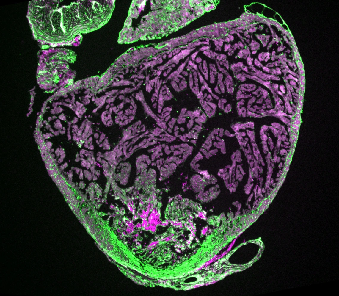
After a heart attack, the human heart cannot replace the muscle it loses. Instead, it forms a permanent scar, which stiffens the heart and can eventually lead to heart failure. Some animals do far better: they can completely rebuild damaged heart muscle. Injury triggers an immune response that unfolds in stages, from fast-acting scavenger cells such as macrophages to slower, more specialised cells such as B cells, best known for producing antibodies. What has remained unclear is which parts of this response tip the balance between scarring and regeneration.
The Mommersteeg Group carries out research using the Mexican cavefish aka Astyanax mexicanus. The same species exists in two forms: a river-dwelling surface fish, which regenerates its heart after injury, and a blind cave-dwelling form from the Pachón cave, which lays down a permanent scar. Using single-cell RNA sequencing, they profiled every cell type in the injured heart of both forms at six timepoints after injury.
Both fish mounted a near-identical immune response in the first three days, but later in surface fish, immune cells stayed in the wound for weeks, while in the cavefish the response faded back towards baseline after day three.
When the team suppressed that late response with an anti-inflammatory drug, the surface fish formed significantly larger scars, showing that the prolonged inflammation, far from being harmful, was necessary. Digging into which immune cells were responsible, they found two mechanisms running one after the other.
The first involves macrophages, the immune system’s scavenger cells. Late-arriving macrophages in surface fish were found to be actively eating debris, and two independent ways of blocking that eating behaviour both left the fish with unresolved fibrotic tissue and much larger scars. It was specifically the macrophages’ specific function, not merely their presence, that mattered.
The second was more surprising. B cells flooded into the wound from seven days after injury in surface fish and were almost absent in the cavefish. Blocking them stopped heart muscle cells from completing cell division, and regeneration failed.
Crucially, the two mechanisms proved independent of one another: removing the macrophages did not stop the B cells arriving, and the two block different steps of repair.
To test whether this was unique to Mexican cavefish, the Bristol-based members of the team looked at zebrafish. B cells accumulated in injured zebrafish hearts in a similar way, and zebrafish genetically lacking mature lymphocytes regenerated poorly, with fewer dividing heart muscle cells, matching the cavefish result.
The findings identify two specific, separable targets for anyone hoping to coax the mammalian heart into repairing itself: clearing scar tissue by phagocytosis and pushing heart muscle cells through cell division. Because the two act independently, tackling both together may work better than either alone.
This research was led by Dr Esra Sengul, Hulme Junior Research Fellow in Medicine at Brasenose College (hosted in the Carr, Mommersteeg, Klemm, and Fernandes Groups) under the supervision of Professor Mathilda Mommersteeg (DPAG) and in collaboration with Professor Rebecca Richardson (University of Bristol).